Scanning Electron Microscopy (SEM) Analysis for Small Molecule Pharmaceutical Characterization

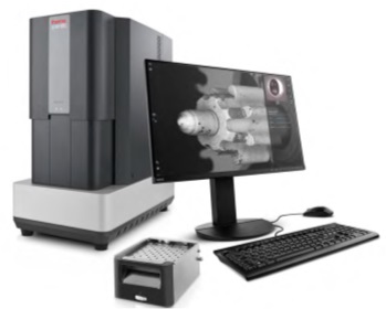
Scanning Electron Microscope Machine

Why is Scanning Electron Microscopy (SEM) Analysis important?

SEM Analysis is an essential technique in pharmaceutical research, providing detailed high-resolution images crucial for the characterization of small molecule drugs. This method is instrumental in understanding and controlling drug formulation and manufacturing processes.

What is SEM Analysis used for?

Our SEM Lab Analysis/SEM Imaging can offer insights into:

  • Particle morphology and size distribution

  • Crystal habit and polymorphic forms

  • Surface defects and impurities

  • Drug-carrier interactions in formulations

  • Coating integrity in drug delivery systems

Scanning electron microscope image

Our SEM Services in Pharmaceutical Small Molecule Analysis

Morphology Assessment

Detailed examination of particle shapes and surfaces, essential for understanding dissolution rates and bioavailability.

Crystal Imaging

Detailed images of the microstructure and surfaces of crystal, information on effective surface area of the crystal.

Polymorph Characterization

Identifying different crystalline forms of a drug, which can significantly impact its therapeutic efficacy and stability.

Our Scanning Electron Microscope (SEM) Process for Pharmaceutical Analysis

1

Sample Preparation:

Careful preparation of the pharmaceutical sample, often involving coating with a conductive layer to enhance imaging quality.

2

High-Resolution Imaging:

Utilizing a focused electron beam to scan the drug particles, achieving nanometer-scale resolution.

3

Signal Detection:

Collecting secondary and backscattered electrons to generate detailed surface topology images.

4

Data Interpretation:

Analyzing SEM images to extract critical information about the pharmaceutical sample’s microstructure and surface characteristics.

Advantages of Our Scanning Electron Microscopy Services for Pharmaceutical Analysis

  1. Cutting-Edge SEM Technology: Utilizing advanced SEM instruments capable of capturing detailed images of small molecule drugs.

  2. Pharmaceutical Expertise: Our team specializes in the analysis of pharmaceuticals, offering deep insights into small molecule characterization.

  3. Tailored Analytical Approaches: Customized studies designed to meet the specific needs of pharmaceutical research and development.

  4. Comprehensive and Compliant Reporting: Providing thorough analysis and reports, compliant with pharmaceutical regulatory standards.

Download Our Free Whitepapers

All FREE Whitepapers are available to download today:

SPA™ Technology Whitepaper

Chemistry Agnostic Whitepaper

UPDATED! Comparison SPA™ Assay vs Shake Flask Whitepaper

SPA™ Assisted Modelling Whitepaper.

Companies Who Have Used Our Services

Frequently Asked Questions

 The main distinction between SEM and TEM lies in how they create images. SEM relies on detecting reflected or dislodged electrons, while TEM utilizes transmitted electrons that pass through the sample to generate images.

When utilizing a scanning electron microscope (SEM), it commonly detects two distinct electron types: backscattered electrons (BSEs) and secondary electrons (SEs).

SEM lets you see compositional differences that optical images miss. When you use benchtop SEM to examine pharmaceutical tablets or powder samples, you’ll notice finer details and compositional differences compared to optical microscopes, even at the same magnification.”


SEM provides a magnification range from approximately 10 to 3,000,000 times, spanning about six orders of magnitude. Unlike optical and transmission electron microscopes, SEM image magnification isn’t reliant on the objective lens power.


Among instrumental methods, the scanning electron microscope (SEM) holds a prominent position for examining and analyzing micro- and nanoparticles alongside characterizing solid objects. SEM’s preference in particle size analysis stems from its remarkable resolution capability, which extends to 10 nm or 100 Å.